What do you really understand about the pelvic floor? It is a bit more discussed these days, but still it is a bit of a mystery to a lot of people.
In this article, Lisa Vernon explains what it is, what it does and why it matters.
Since working in the field of womens health physiotherapy, it has become clear to me that many women have a limited understanding of what the pelvic floor muscles are, where they are and what the role of the pelvic ligaments and muscles is.
In fact, before I started studying Womens Health Physio and the in-depth learning of the Level 1 Women’s Health course through the Australian Physiotherapy Association, I could include myself in that assessment!
I hope to share some of this information and help expand your understanding in this article.
A pelvic floor analogy
A nice analogy to understand the pelvic floor is to think of a ship in the docks.
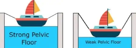
The dock represents the bony structure of the pelvis.
The ship represents the pelvic organs – that is the bladder, uterus and rectum in a female.
It is worth mentioning that males have a pelvic floor also, but this discussion is in relation to female pelvic health and function.
The ropes represent the ligaments and fascia (connective tissue) that help to suspend the organs and keep them in their correct anatomical position. You could say that the ropes help to maintain a ship in the right position in relation to the dock.
The water in the dock represents the pelvic floor muscles and the upward support they provide to the organs, and therefore the suport that they supply to ligaments and fascia.
If the muscles are strong, they will sit higher and support the pelvic organs from below.
If the muscles are weak, from inactivity or following trauma (post childbirth), the pelvic organs will sit lower.
The pelvic organs place more load on the ligaments which will elongate over time, which creates a higher risk of pelvic organ prolapse.
So, what exactly is the pelvic floor??
The pelvic floor is not just pelvic floor muscles!
In 2017, the International Urogynaecological Association and International Continence Society published their terminology report stating that:
The ‘pelvic floor’ are the structures located within the bony pelvis ie urogenital and anorectal viscera, pelvic floor muscles and their connective tissues, and nerves and blood vessels.
Urogenital and anorectal viscera are the pelvic organs – that is the bladder, uterus and rectum in a female. Viscera is another name for organs.
Therefore the terms pelvic floor and pelvic floor muscles are not interchangeable.
So when pelvic floor function and dysfunction is discussed it does not just refer to the pelvic floor muscle function and dysfunction.
Pelvic floor dysfunction also includes
bladder and bowel dysfunction, pelvic organ prolapse (POP), sexual dysfunction and pain.
What makes up the pelvic floor?
Bony pelvis
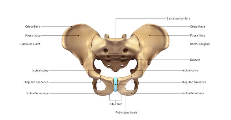
The pelvis consists of 3 bones – a hemi-pelvis on either side and the sacrum at the back.
This forms a solid bony ring that contains the pelvic organs and the pelvic floor, as well as allow passage of key vessels and nerve branches and lymphatics to the lower limbs.
The top edge of the ring is called the pelvic brim or pelvic inlet. Anything below this pelvic rim is said to sit within the pelvic cavity.
The pelvic organs
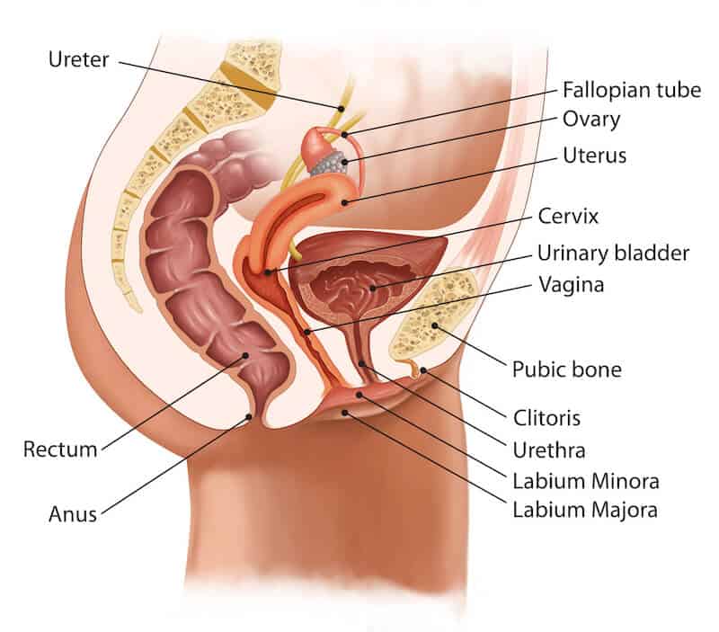
Within the female pelvis are the
-
- The female reproductive system
-
- The lower urinary tract
-
- The lower digestive tract
Each of these pelvic systems and tracts has 2 structures, one for storage, and one for release
| Lower urinary tract | Reproductive system | Digestive tract | |
| Storage | Bladder | Uterus | Rectum |
| Release | urethra | vagina | Anal canal |
As you can see in the diagram, the urinary structures sit in front of the structures of the reproductive system
Therefore the posterior (back) aspect of the bladder and urethra rests next to the anterior (front) of the vagina, and the anterior (front) aspect of the rectum and anal canal rests towards the posterior (back) wall of the vagina
Ligaments and fascia
Fascia is strong and sometimes stretchy tissue that covers, separates, or holds together different organs, muscles, blood vessels, and nerves.
The fascia within the pelvis is known as the endopelvic fascia, which translates to fascia within the pelvis
This fascia runs between and around the pelvic organs and serves to suspend the organs from above and to hold the pelvic organs in the correct anatomical place
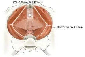
The rectovaginal fascia occupies the space between the rectum and the vagina. The fascia prevents the rectum from bulging forward into the vagina. Women with rectoceles (rectum bulging forward into the vagina) have defects in this fascia. This is what is repaired in women with posterior vaginal wall prolapse.
The pubocervical fascia separates the bladder and urethra from the anterior vaginal wall. Women with cystoceles (bladder bulging into the vagina) have defects in this fascia. This is what is repaired in women with anterior vaginal wall prolapse. In women with stress urinary incontinence the same fascia may be weakened, resulting in a lack of support for the upper urethra.
The uterosacral and cardinal ligaments attach to the cervix and support/ stabilise the uterus within the pelvic cavity.
The cardinal ligaments stabilise laterally (from the side) while the uterosacral ligaments run from the sacrum (tailbone) to the cervix providing support from front to back.
Women with enteroceles, vault prolapse or uterine prolapse (descent of the uterus into the vagina) have defects in uterosacral and / or cardinal ligaments.
Muscles of the pelvic floor
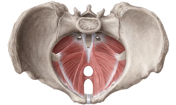
The pelvic floor muscles are at the base of the pelvis, also known as the pelvic outlet.
When they contract, the pelvic floor muscles offer support to the pelvic organs (the water level in the analogy above), thereby reducing tension on the ligaments and fascia (the ropes)
There are 2 layers of pelvic floor muscles
Deep pelvic floor layer
These muscles provide upward support of the pelvic organs and maintain the anorectal angle
-
- Levator ani and coccygeus
-
- Levator ani translates to lift the anus.
Superficial pelvic floor
These muscles provide added closure to the entrance ways of the vagina and anal canal
-
- urogenital triangle muscles at the front, which assist closure pressure around the urethra and vagina.
-
- External anal sphincter which surrounds the anus. An intact and functioning anal sphincter is important in maintaining faecal continence.
Summary
The tems ‘pelvic floor’ and ‘pelvic floor muscles’ are used interchangeably at times, but strictly speaking, the pelvic floor muscles are just one component of the pelvic floor.
A lot of women don’t understand the basic anatomy of the pelvic floor.
A basic level of knowledge is worthwhile to understand the roles of the pelvic floor muscles and how they contribute to supporting the pelvic organs and helping maintain continence.
It also helps to see them in action!
Lisa Vernon can help you do that with the aid of Real-Time Ultrasound imaging.
This can make up part of her Womens’ Health Physiotherapy assessment process.
Interested in finding out more about what a Womens Health Physio appointment looks like? Got some questions? Check out this page which should answer a lot of them!
Reference
Bo et al – An International Urogynaecological Association (IUGA/international Continence Society (ICS) joint report on the terminology for the conserviative and nonpharmalogical management of female pelvic floor dysfunction. International Urogynecology Journal. 2017 Feb 1:28(2): 191-213