
If this sounds like you, Real Time Ultrasound might be what you are looking for!
Real Time Ultrasound (RTUS) is a really valuable tool that provides a ‘window’ into the body in a non-invasive way.
It can be used to see lots of things – muscle, tendon, bladder volume and of course, babies!
At Adelaide West Physio and Pilates, we use RTUS to allow both the patient and the physiotherapist to view a muscle contraction as it happens.
Lisa Vernon, our Women’s Health Physio, uses this frequently as a non-invasive way of assessing the pelvic floor as part of her Women’s Health Service.
This is how we use RTUS at Adelaide West:
This visual feedback can be helpful to learn how to make a muscle work more or less. It can also be used to learn how to make a muscle work earlier or later in function.
If you are trying to learn how to activate a muscle that is deeply placed in the body and not visible from the outside of the body, this kind of feedback can be very useful.
RTUS can be used for any muscle. With physios, it is most commonly used for learning or improving how to contract the deep abdominal muscles, like transversus abdominis, multifidus and the pelvic floor.
Recently, we have found it to be even more useful for learning how NOT to contract certain muscles, like overrecruiting outer abdominals and learning how to relax the pelvic floor muscles.
It is great to show patients that they don’t have to brace with everything they have. Instead it is a case of ‘not too little – not too much’.
RTUS can be helpful in the following situations:
If you are doing Pilates, the Real Time Ultrasound assessment may be a part of your initial assessment, as part of the process of learning how to feel your deep stabilising or ‘core’ muscles.
To get an image of transversus abdominis, you are lying on your back or your side with your tummy exposed, and the probe is put just above your hip bone on your side.
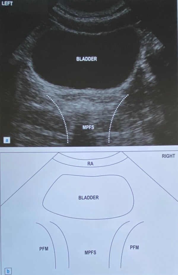
To get an image of your pelvic floor, you are lying on your back too, and the probe is put on your tummy and angled down in the direction of your pubic bone to assess pelvic floor function or effectiveness of pelvic floor exercises.
To get an image of multifidus, you are lying on your tummy and the probe is put on your back muscles.